Clearly bisect the carina or bronchi. The tube is inserted by the use of a guide wire called the stylet see image1 which removed after the tube correct placement is confirmed.

Iatrogenic Bronchopleural Fistula From A Dobhoff Tube Radiology Case Radiopaedia Org
The Dobhoff tube was introduced in the mid-1970s by surgeons Robert Dobbie and Jim.

. It is also important to note that feeding tube insertions causing pulmonary complications are not always related to tracheobronchial insertion. They will then insert the Dobhoff tube through the nose into the stomach and into the duodenum. ZWith NG tubes placement should be obvious.
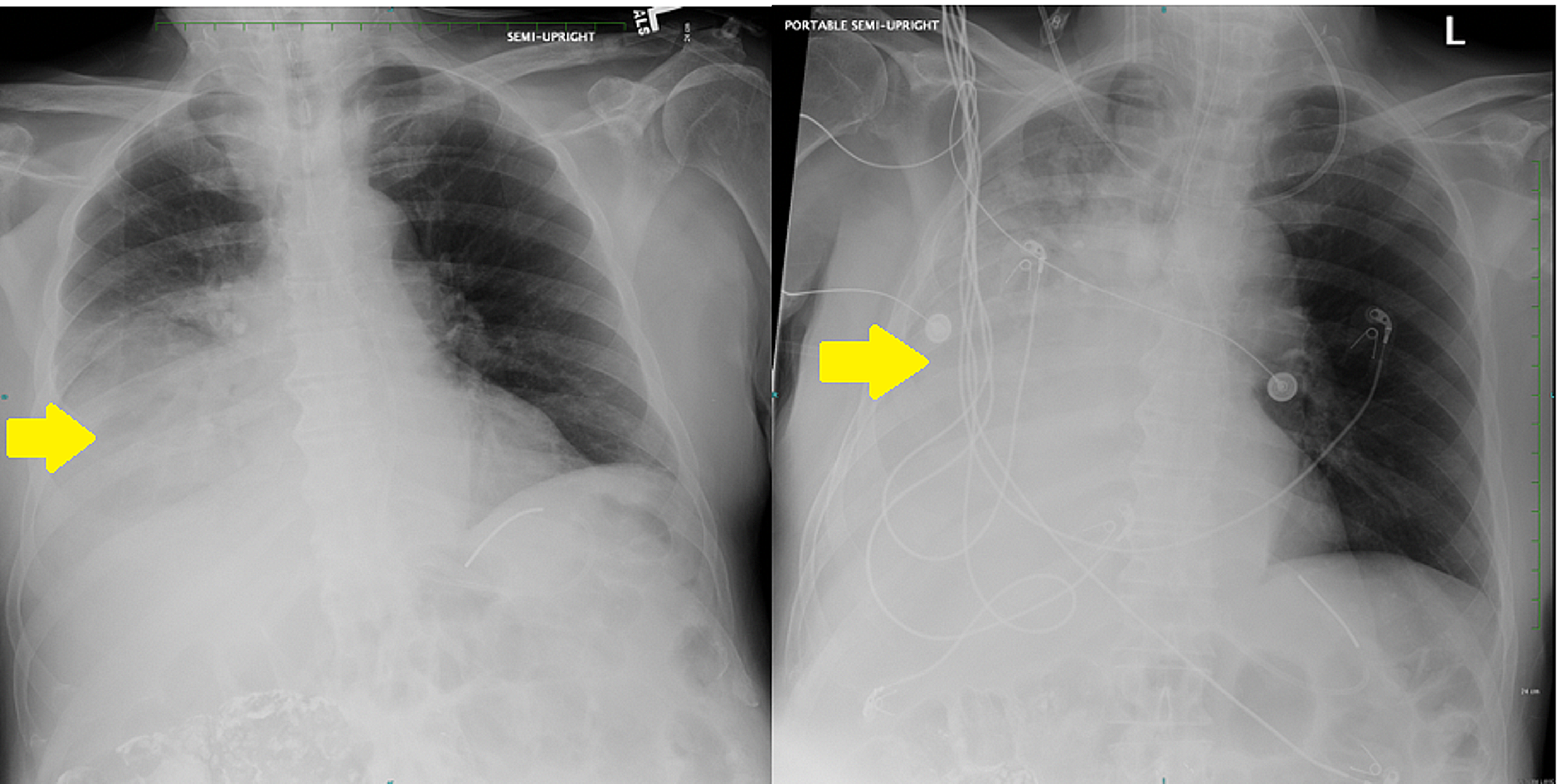
After insertion correct placement in the body is usually checked with the help of X-rays or fluoroscopy. Most tubes are visible on a chest x-ray without a guide wire. A review of the x-ray showed that the feeding tube was in the main bronchus.
We had a situation where a dubhoff did not show up anywhere on the x-ray. Tube feedings were begun. Most however are placed in the stomach.
Cross the diaphragm in the midline. Data was collected at initial placement prior to x-ray confirmation. Descend in the midline following the path of the esophagus and avoiding the contours of the bronchi.
When a Cortrak machine is not available a two step technique using a chest x-ray to confirm placement in the esophagus and then through the pylorus is an effective way to prevent serious complications23 rEFErEncES 1. Auscultation was performed in all 78. Follow-up abdominal x-ray revealed displacement of the Dobhoff tube in the left pleural space.
A Dobhoff or any type of weighted tube placed by stylet cannot be checked for placement via air burp method. In order to prepare a patient for the insertion of a Dobhoff tube the esophagus and nasal cavity are numbed and the patient if conscious may be given a mild sedative. The wire was left in.
Tube crosses the diaphragm in the midline. It was a 12 french by the way. Nasopharynx with 2 Lidocaine jel ly.
You need to be confident that you can see the tip. An x-ray can ensure that the Dobhoff tube has been placed correctly. Aspiration and a Dobhoff tube was subsequently placed at the bedside for delivery of enteral nutrition.
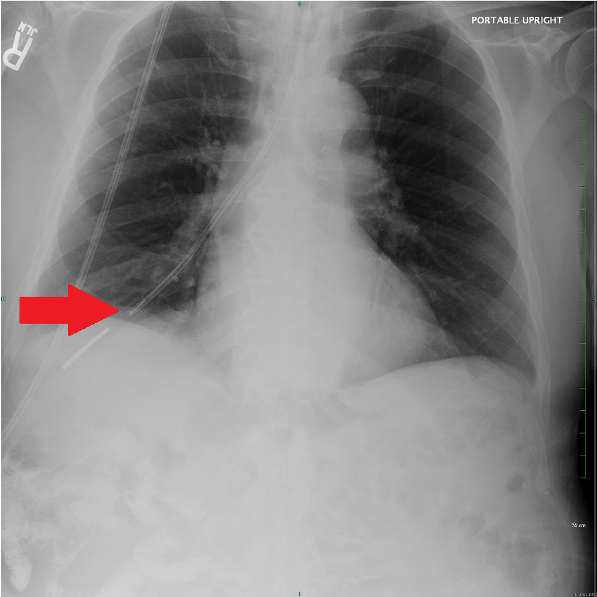
Once the tube is in the proper place Gastroview contrast is injected into the tube and an image is taken to document the placement. Therefore bilateral chest tubes were placed. A chest X-ray performed shortly after the tube placement demonstrated that the tip of the Dobhoff tube was within the right lung base following the course of the right mainstem bronchus Figure 1.
The x-ray was read and placement confirmed. Most tube positions are checked by assessing pH of tube aspirate. The patient experienced respiratory distress.
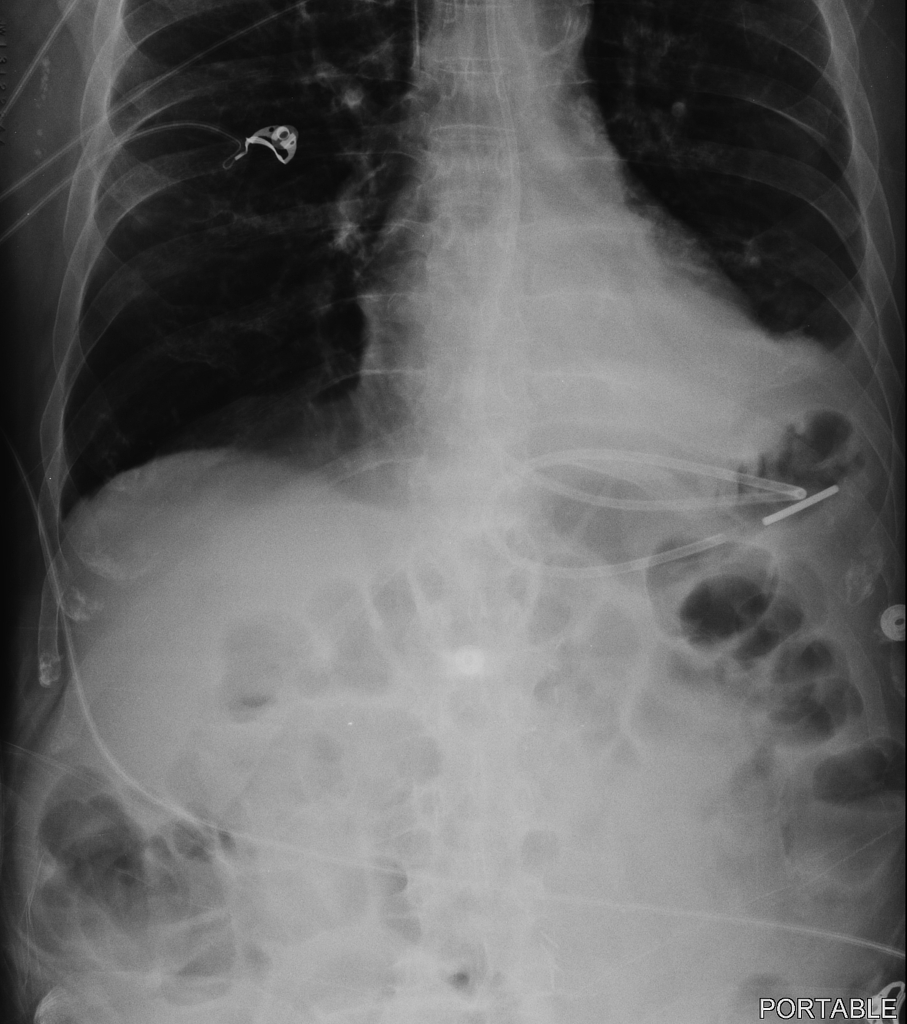
I am pretty sure the only way to verify placement of a dobbhoff is by x-ray since it actually goes into the duodenum. Tube feedings were initiated. Abdominal X-ray after Dobhoff tube DHT placement to confirm accurate positioning.
An abdominal X-ray was obtained to confirm placement of the DHT Figure 1. It must be checked via Xray then we would place the patient on their left side for a while to allow natural peristalsis to advance it further if needed. The tip sits below the diaphragm.
At our facility we x-ray all feeding tubes for placement verification. Steps for NG Feeding Tube Placement in an Awake Patient. Measure tube from tip of nose to subxyphoidprocess about 3035cm in most patients Step 2.
Of 78 nasoenteral intubations in 46 patients using a Dobbhoff Biosearch Medical Products weighted enteral feeding tube gastric aspirates were evaluated in 28. A nasal bandage is used to secure the tube. ZEndotracheal placement zEpistaxis zSinusitis.
The patient was found dead. However in our patient who had history of Roux-en-Y the DHT bypassed the duodenum and. Dobhoff tubes come with a radiopaque stripe making them easily visible in X-rays.
Normally the DHT tip should be placed in the 2 nd to 3 rd portion of the duodenum and would create a C-shaped tracing on the X-ray. Potential Complications Some complications that could occur during the insertion of a Dobhoff tube include. A Dobhoff tube was placed by a house physician.
After removal of the tube a follow-up chest x-ray revealed iatrogenic bilateral pneumothoraces. When tube is connected to low intermittent suction there should be return of gastric contents If there isnt confirm placement with xray KUB zWith dobhoff tubes should always confirm placement as no suction will be applied. Patients are usually positioned on the right side while the tube is put into the nose.
Dobhoff tube is a special type of nasogastric tube NGT which is a small-bore and flexible so it is more comfortable for the patient than the usual NGT. Tube bisects the carina. The side hole is usually located just proximal to the tip.
Tip of feeding tube should be in 2nd or 3rd portion of duodenum. The nurse is sure of gastric placement. Acute hypoxemic respiratory failure ensued.
1 1 1 Open Access Case. Tube descends the thorax in the midline. Have its tip visible below the left hemidiaphragm.
We told the nurse to check by air bolus while we await a second xray to be completed and read. Due to the limitations of bedside techniques in confirming Dobhoff tube placement x-ray remains the gold standard in confirmatory testing 1910. The distal tip of the feeding tube is in a loop of jejunum in patient status post gastrojejunostomy.
Entering a bronchial tube. A correctly placed nasogastric tube should 10. The physician confirmed placement after reading the x-ray.
Dobhoff tube is an excellent alternative although it can carry a serious risk of pneumothorax if placed blindly. Place tube through nares and ask patient to swallow as you pass the tube. What can go wrong.
Placement of the tube is checked by a post-insertion radiograph centered on the. Feeding tube with guidewire brown tip that is 120cm preferred over blue tip dobhoff tube Lubricant 60 ml syringe. I was also taught that you should keep the guide wire handy since it may be need to replace the tube.
A DHT was inserted after intubation for feeding purposes.

Abdominal X Ray Revealing The Dobhoff Tube Traversing The Left Main Download Scientific Diagram
J2kuu2zup1vbbm
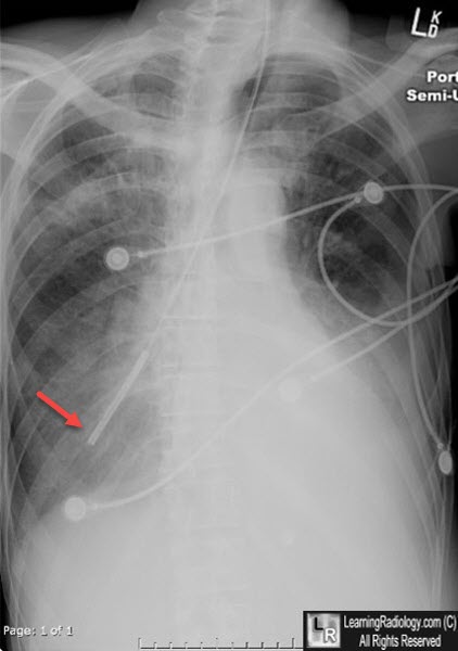
Learningradiology Dobhoff Dobbhoff Tube Malplaced Rll
Dobhoff Nasogastric Tube Tube Radiology Case Radiopaedia Org

Abdominal X Ray After Fluoroscopic Guided Dobhoff Tube Placement Small Download Scientific Diagram

Icu Chest Radiography Lines Ng Dobhoff Etc Youtube
Cureus Hemothorax Following Traumatic Dobhoff Tube Insertion

Learningradiology Dobhoff Dobbhoff Tube Malplaced Rll
0 comments
Post a Comment